
General information
The Sternocleidomastoid is a paired muscle within the superficial layers out of the anterior portion of the neck. It is one of the largest and most superficial cervical muscles.
Literal meaning
The clavicle shape or form.
Interesting information
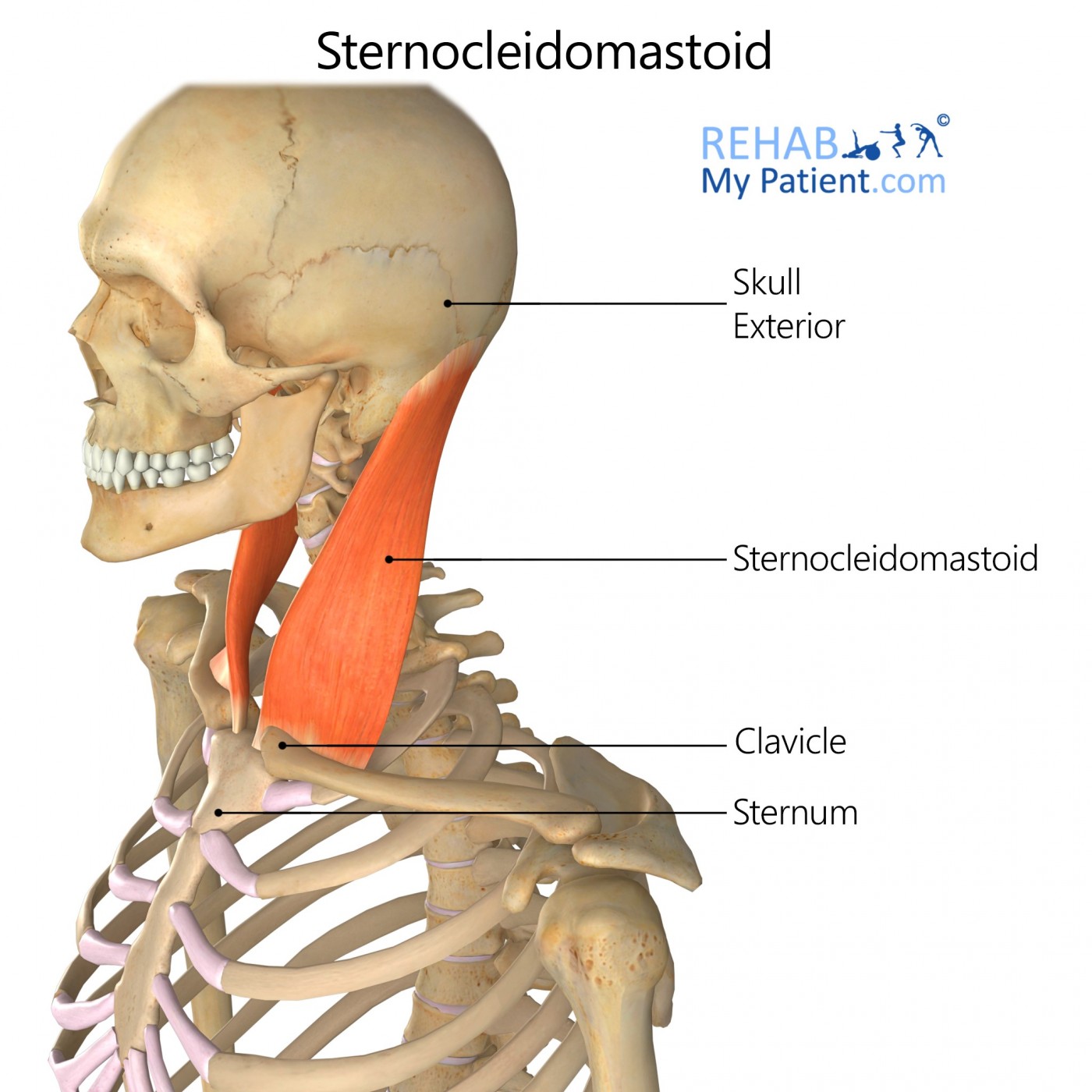
The sternocleidomastoid muscle is found at the base of the sternum and attaches itself to the clavicle and the mastoid process for the temporal bone on the base of the skull. One of the muscles is located on either side of the neck and the muscle controls lateral flexion, which handles the rotation and tilting of the head from left to right. Sudden movements such as whiplash can cause the muscle to become injured.
Strained SCM muscles may cause swelling and redness around the injury site. In a severe case, the skin may become discoloured or bruised along the path where the injury occurred. If the strain results in a spasm, twitching or fluttering beneath the surface will occur. Apply ice for 20 minutes at a time to help relieve the redness and swelling.
Origin
Anterior upper margin out of the manubrium of the sternum.
Superior aspect from the medial third of the clavicle.
Insertion
Lateral side of the mastoid process and the lateral portion of the superior nuchal line.
Function
Extension of the head to rotate it to the opposite shoulder unilaterally.
Flexion and extension of the head.
Raises the rib cage.
Nerve supply
Accessory nerve.
Ventral rami of the cervical spinal nerves C2-C4.
Blood supply
Occipital branch out of the posterior auricular from superior to inferior.
Occipital artery’s sternocleidomastoid branches.
Superior thyroid artery of the sternocleidomastoid artery.
Suprascapular and superficial cervical arteries of the subclavian artery.
Relevant research
The study sought to describe the 3-dimensional organization of any connective tissues within the suboccipital region. A sectional anatomic investigation using E12 sheet plastination was conducted. Six human cadavers, two of which were male and four were female, ranging in age from 54 to 86, were used to complete this study. Five of the participants were sectioned in 2.5-mm-thick coronal, sagittal or transverse sections. There was no visual aggregation of the fibrous connective tissue between the trapezius and the sternocleidomastoid muscles. The intervening space was occupied fully with fatty tissue that was not distinguishable from the subcutaneous tissue.
Zhang M, Lee AS. The investing layer of the deep cervical fascia does not exist between the sternocleidomastoid and trapezius muscles. Otolaryngol Head Neck Surg. 2002;127(5):452?454. doi:10.1067/mhn.2002.129823
Sternocleidomastoid exercises
Lateral flexion
Lateral flexion is a motion that involves tipping the head to one side. The exercise simulates the motion and it is performed sitting on a weight bench with the body in a side lying position. Begin on the right side with the legs stacked and the head just past the bench’s end with the right hand on the floor to brace the body. Carefully holding a weight plate on the left of the head, bend the head slowly down. Lift the head as high as possible and hold for just a second. Complete 10 repetitions before switching sides. Make sure the body is as still as possible.
Sign Up
Sign up for your free trial now!
Get started with Rehab My Patient today and revolutionize your exercise prescription process for effective rehabilitation.
Start Your 14-Day Free Trial