
General information
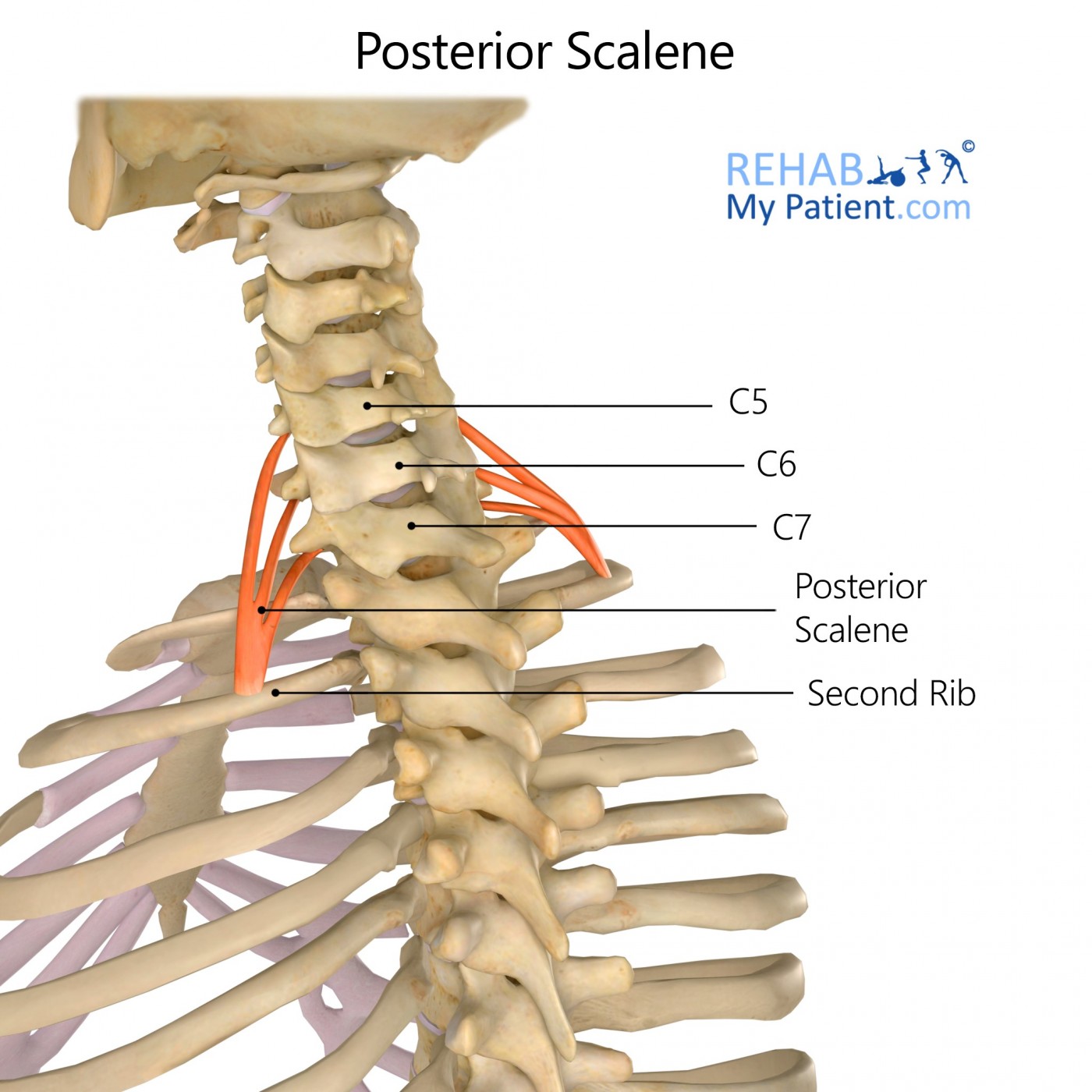
Posterior scalene is a lateral muscle on the inferior half of the neck.
Literal meaning
Muscle with three unequal sides that is behind.
Interesting information
Posterior scalene is one pair of the group of three pairs of scalene muscles. Posterior scalene elevates the second rib and tilts the neck to the same side. Injury often occurs to the group of scalene muscles due to whiplash injuries, excessive coughing, pulling and lifting, and carrying a heavy backpack or handbag. Many times, patients report pain in other areas outside of the Posterior scalene area, but the pain is often directly attributed to this muscle without them being aware of it. Treatment can include moist heating pads, proper ergonomic work habits, ibuprofen, massages, and physical therapy.
Origin
Posterior tubercles of transverse processes of fourth to sixth cervical vertebrae.
Insertion
Lateral surface of second rib posterior to the tubercle for the serratus anterior.
Function
Laterally flexes the lower cervical vertebrae.
Elevates the second rib.
Nerve supply
Cervical and brachial plexuses.
Blood supply
Ascending cervical and various muscular branches from the inferior thyroid.
Suprascapular artery, the superficial cervical artery, and the first posterior intercostal artery.
Relevant research
This case study presents an obscure and often overlooked cause of shoulder pain: Neurogenic Thoracic Outlet Syndrome (NTOS). A typical case of NTOS presents with symptom of a nagging pain that increases with exercise and persists after cessation of activity. The pain is often present at rest. Patients with NTOS typically undergo diagnostic arthroscopy, during which an interscalene or other brachial plexus block is performed.
Boezaart, A. P., Haller, A., Laduzenski, S., Koyyalamudi, V. B., Ihnatsenka, B., & Wright, T. (2010). Neurogenic thoracic outlet syndrome: A case report and review of the literature. International journal of shoulder surgery, 4(2), 27.
The thoracic outlet is divided into three tunnels: the interscalene triangle, the costoclavicular space, and the retropectoralis minor space. Compression or irritation in this area can result in thoracic outlet syndrome (TOS). Magnetic resonance (MR) imaging is useful for assessing the thoracic outlet and the brachial plexus.
Demondion, X., Boutry, N., Drizenko, A., Paul, C., Francke, J. P., & Cotten, A. (2000). Thoracic outlet: anatomic correlation with MR imaging. American Journal of Roentgenology, 175(2), 417-422.
Posterior scalene exercises
Scalene stretch #1
Sit in a secure and flat-surfaced chair. With the left hand, either grab the underside of the chair seat or place it under the glutes. This positions the shoulder to stay down, preventing the shoulder from raising during the stretch. Tilt the head directly to the right, bringing the right ear towards right shoulder. Bring the right hand up and lightly pull head down towards the right, lightly pressing the stretch further. Hold for 10 counts. The stretch may be repeated with the addition of slightly tilting the head in front of and behind the shoulder line. Repeat the exercise on the left side.

Scalene stretch #2
Sit forward on the edge of a chair, with the chest up. Hold the seat of the chair tightly with one hand to keep ribs down. Side flex head to the opposite side without flexing your head forward. Rotate head slightly toward hand holding chair and slightly extend neck to get more of a stretch.
Sign Up
Sign up for your free trial now!
Get started with Rehab My Patient today and revolutionize your exercise prescription process for effective rehabilitation.
Start Your 14-Day Free Trial