
General nformation
The plantaris muscle is often disregarded as a small, functionless muscle. It is approximately 2-4 inches in length comprised of a thin elongated tendon and a slender muscle belly. Though often deemed unimportant, it is one of three plantar flexor muscles in the hindmost compartment of the leg. The other two are the gastrocnemius, and soleus.
Literal meaning
The name plantaris translates literally as “pertaining to the sole of the foot”.
Interesting information
Because of its minimal functionality, the plantaris is most commonly used for reconstruction or tendon grafting in other regions of the body.
It is sometimes mistaken for a nerve and is referred to as the “freshman nerve”.
It is absent in approximately 7-10% of the human population.
Origin
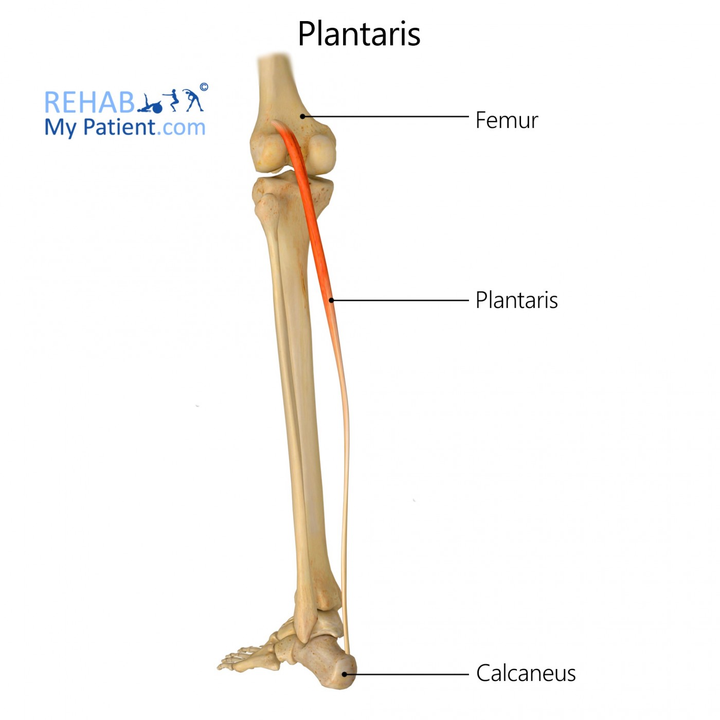
It arises from the lower part of the lateral supracondylar line of the femur in close proximity to the source of the lateral head of gastrocnemius.
It may originate from the oblique popliteal ligament.
Insertion
Passing posteriorly by the lower central region of the knee joint, it takes on a tendon-like structure as it passes distally to implant into the calcaneal (Achilles) tendon of the posterior leg.
Function
The muscle scarcely aids in the following:
• Plantarflexion of the ankle joint causing the foot to move away from the shin
• Flexion of the knee joint
Additionally, it may aid in the provision of proprioceptive data to the central nervous system concerning the placement of the foot.
Nerve supply
Tibial nerve (S1, S2).
Blood supply
Sural arteries.
Relevant research
Despite the controversy, it has now been accepted that injury of the plantaris may occur in isolation. Because symptoms of these injuries sometimes mirror those of other more serious conditions such as DVT (deep vein thrombosis) and gastrocnemius tears, accurate diagnosis by way of ultrasonic and or MR imaging is essential. The most severe plantaris injury is a rupture at the musculotendinous junction but injury most frequently occurs midway the calf.
Leekam, R. N., Agur, A. M., & McKee, N. H. (1999). Using sonography to diagnose injury of plantaris muscles and tendons. AJR. American journal of roentgenology, 172(1), 185-189..
Plantaris exercises
Jumping rope is a fun way of exercising the muscles of the calf and in particular the plantaris muscle. Start by jumping the rope slowly and ensure to land on the ball or your feet rather than flatly onto the floor. After 3 minutes, increase your pace to create a more intense work out. Continue for ten minutes then gradually slow down before coming to a halt. This may be repeated with short breaks between each set.
Another easy option is to sit upright on a chair with both knees bent and feet planted flat on the floor in front of you. Now press your toes firmly into the ground and raise your heels upward. Hold this position for ten seconds before placing the feet back to their original position. Repeat 30 times. Note that you may alternate each foot or exercise both simultaneously.

In the event that there has been injury to the plantaris muscle, it is advised that little pressure be exerted on the muscle in the one week period following injury. Rest, icing and elevation of the injured leg are the most common suggestions given to patients. As time passes however, you may engage in light stretches which will result in distension of the developing scar over the wounded muscle and stimulate collagen growth, thereby improving recovery time.
Sign Up
Sign up for your free trial now!
Get started with Rehab My Patient today and revolutionize your exercise prescription process for effective rehabilitation.
Start Your 14-Day Free Trial