
The knee joint is referred to as a synovial joint. This joint forms a connection between two bones that together make a moving articulation. At the end of the bones, a joint surface is lined with a low-friction material known as articular cartilage. The cartilage is lubricated from a film of fluid that is referred to as synovial fluid. This fluid is produced from an extremely thin lining known as the synovium. It is supported from a stronger fibrous structure containing the joint known as the joint capsule.
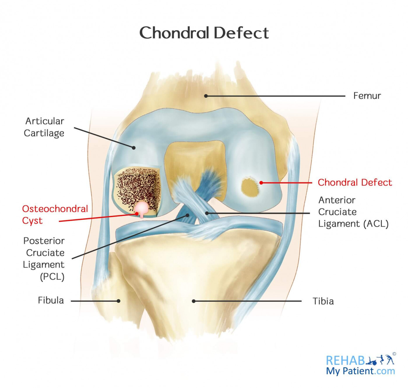
Any portion of the joint is prone to injury. A common injury to the knee is a localized loss of cartilage, referred to as the chondral defect, which combines with an injury to the supporting bone.
A chondral defect can also lead to a small cyst in the top of the bone, just under the cartilage, as a result of repetitive trauma to the knee. They typically occur in the upper part of the tibia.
Chondral Defect Anatomy
The knee is one of the most complex and largest joints within the body. It joins the shinbone (tibia) and the thighbone (femur). The smaller bone running along the tibia is called the fibula, and the kneecap (patella) are the other bones making up the knee joint. Tendons keep the knee bones connected with the leg muscles that are responsible for moving the knee joint. Ligaments keep the knee bones joined together and deliver stability to the knee.
The lateral and medial menisci are two c-shaped cartilage pieces that serve as shock absorbers between the tibia and the femur. Countless fluid-filled sacs help the knee to move freely.
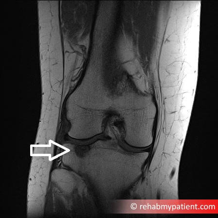
Chondral defects are almost always diagnosed after a magnetic resonance imaging (MRI) scan. They are not possible to be detected by way of physical examination, but sometimes the clinical history can point towards a chondral defect being the cause of the pain. However, 99% of the time, you will need an MRI to diagnose this condition.
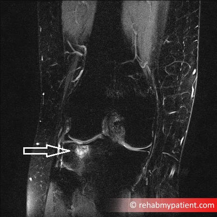
An MRI scan showing a chondral defect with bone bruising
How to Treat a Chondral Defect:
- Stop aggravating the problem
If you continue to play sport, or put heavy weight bearing through the joint, then you may not recover. Chrondral defects can take a long time and are often associated with some bone bruising, and resting the knee from sport can provide significant relief over a longer term. But you must be patient with it.
- Arthroscopy
If a partial thickness injury to the cartilage and the mechanical symptoms for intermittent and catching pain are present, arthroscopy might be necessary to smooth the surface of the joint and remove any loose cartilage. Also the defect can be drilled slightly which can fill the area with blood and stimulate healing (see microfracture below).
- Internal Fixation
In certain instances, the damaged cartilage area and the underlying bone will stay attached partially or they can be loose but still in one piece in the joint. Depending on the situation, fragment fixation back to the defect is desirable. This is a rare form of treatment.
- Microfracture
If a full thickness of cartilage is detected, the aim is to restore the cartilage that covers the bone underneath. Articular cartilage doesn’t have a blood supply, so it doesn’t possess the ability to heal when damaged. A microfracture operation is completed along with keyhole surgery where they make multiple small holes within the bony base of the defect that allows the blood to enter into the cavity and start the healing process. Post-operative treatment is imperative to the success of the procedure.
Tips:
- Most often chrondral defects are caused by repetitive trauma, for example, skiers or rugby players who put their knees through a lot of stress.
- Occasionally other more significant accidents can cause it, such as a motor vehicle accident when blunt force trauma occurs to the knee.
- Sports injuries leave an individual open to a number of different injuries that take an extended period of time to recover from.
- For those who partake in lifting weights, you need to make sure you perform the movement correctly to prevent injuries to the knee.
- Stepping onto an uneven surface can cause problems with the knee and create a chondral defect.
- Running on surfaces that are uneven can cause undue pressure and strain on the knee muscles and joints.
Sign Up
Sign up for your free trial now!
Get started with Rehab My Patient today and revolutionize your exercise prescription process for effective rehabilitation.
Start Your 14-Day Free Trial