
General information
The rectus capitis anterior muscle is a flat, short muscle that is nestled behind the upper portion of the longus capitis.
Literal meaning
Straight muscle of the back of the head.
Interesting information
Nestled deep between the first two of the cervical vertebrae and the skull base, these muscles help to maintain alignment between the upper cervical vertebrae and occiput. It helps to support proper functioning of the associated circulatory and nervous structures. Suboccipitals are also responsible for generating the fine movements for the head, such as when reading a book or scanning the roadways when driving or walking. These movements are important when maintaining awareness and orientation of the head while the body is moving.
Turning too far to one side or too quickly can cause injuries to this muscle. Whiplash tends to be one of the most common methods for injury the muscles in the neck. Symptoms include pain in the immediate and radiating area and headaches. Common treatments include isolating movement of the area and using anti-inflammatory medications to reduce pain and swelling.
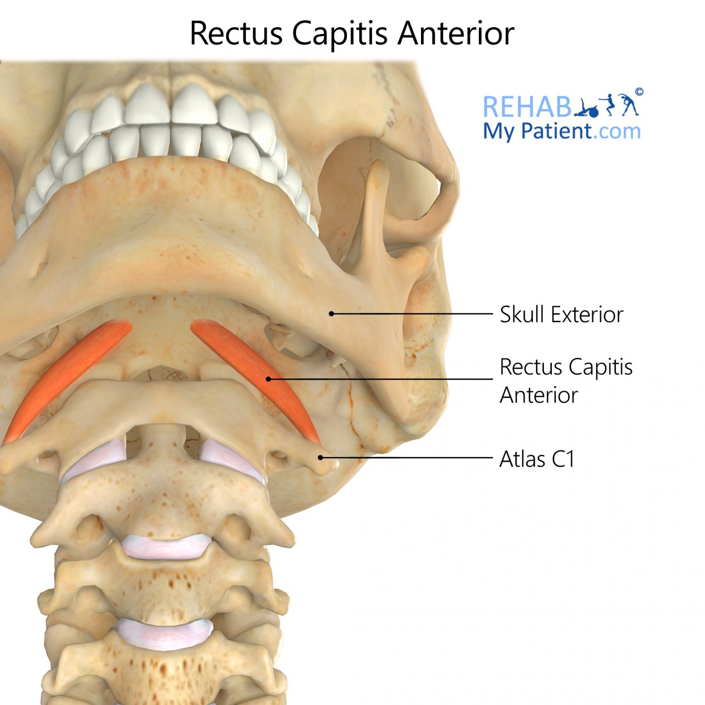
Origin
Anterior margin for the lateral mass of the atlas located at the base of transverse process.
Insertion
Interior surface of occipital bone located anterior to occipital condyle.
Function
Weakly assists with the head’s flexion.
Postural muscle helping to monitor the position of the head and movement.
Nerve supply
Branches from the cervical rami C1-C2.
Blood supply
Ascending cervical artery supplies the muscle.
Relevant research
Studies were conducted into the underlying pain associated with injuries from whiplash. They indicate that the injuries produced a plasticity of changes in the various neuronal structures responsible for amplification of exaggerated and nociception responses to pain. Evidence is consistent for the central nervous system’s hypersensitivity to any sensory stimulation when faced with chronic pain from whiplash injuries. Damage to the tissues, whether visible or not, is probably the main component to central hypersensitivity.
Various mechanisms underlie and co-exist for those faced with chronic whiplash. Patients suffering from chronic pain after the injury who have spinal cord hyperexcitability will often be in an extreme amount of pain following any type of stimulation. When spinal hypersensitivity exists, it may explain the pain when there is no tissue damage detected. Whiplash is an intense condition where some people show neuropathic pain. Predominant neuropathic pain relates to a higher degree of pain and disability.
Davis, Charles, G., (February 2013). “Mechanisms of Chronic Pain from Whiplash Injury.” Vol. 20, Is. 2, 74-85.
Rectus capitis anterior exercises
Suboccipital stretch
Stand with the back and head against a wall, or sit with the back pressed into the back of a chair. Pull the chin back until the neck is straight. If performing the exercise against the wall, pull the chin back until the neck is touching the wall. Hold for five seconds before you release. Don’t lift the chin upward for this exercise. Perform the exercise in repetitions of ten, up to seven times during the course of the day.
Sign Up
Sign up for your free trial now!
Get started with Rehab My Patient today and revolutionize your exercise prescription process for effective rehabilitation.
Start Your 14-Day Free Trial