
General information
Occipitalis is often referred to as the occipital belly of the epicranius muscle.
Literal meaning
The muscle on the back of the skull.
Interesting information
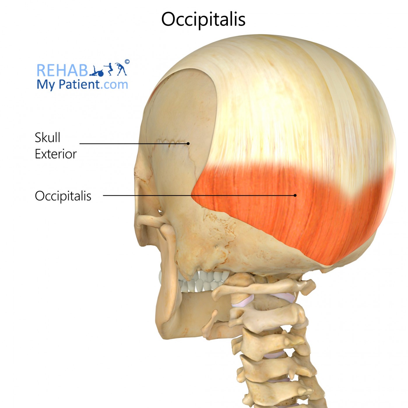
The occipitalis muscle, or occipital belly, is a muscle situated on the back of the skull. Some anatomists consider occipitalis and frontalis to be two discrete muscles while others prefer to categorize them as two regions of the same muscle unit – the epicranius, or occipitofrontalis.
Occipitalis is a relatively thin, rectangular shaped muscle which arises from the occipital bone and inserts in the epicranial aponeurosis. Its primary function is to move the scalp towards the posterior of the skull.
Occipitalis works with frontalis to manipulate the scalp which aids in the formation of human facial expressions by raising the eyebrows and creasing the forehead. Therefore, serious injury or nerve damage to occipitalis may present as decreased movement of the scalp.
Apes also possess an occipitofrontalis muscle, but for them it serves a greater purpose than mere facial expression. It aids in balancing the head on the vertebral column by pulling back on the skull. This extra physiological function of occipitofrontalis in apes is due to the fact that they are not bipedal like humans.
Origin
Highest nuchal lines.
Insertion
Epicranial aponeurosis (posterior surface).
Function
Pulls the epicranial aponeurosis posteriorly toward the occipital bone; works in conjunction with the frontal belly of epicranius.
Nerve supply
Posterior auricular nerve (facial nerve).
Blood supply
Occipital artery.
Relevant research
Correct diagnosis of injury or other pathology in the scalp requires that radiologists and other health professionals be able to accurately interpret imaging of the scalp including the galea aponeurotica and muscular structures like occipitalis. Computed tomography, magnetic resonance imaging (MRI), and ultrasound are imaging techniques that are applicable to the scalp.
Sharman AM, Kirmi O, Anslow P. (2009). “Imaging of the skin, subcutis, and galea aponeurotica”. Semin Ultrasound CT MR. 30(6):452-64.
Cervicogenic headaches present as unilateral headache pain which radiates from the neck up to supra-orbital region of the skull. When conventional treatments such as pharmaceuticals and physiotherapy are unable to yield satisfactory pain relief, direct injection of the nervus occipitalis major may be required.
Van Suijlekom H, Van Zundert J, Narouze S, van Kleef M, Mekhail N. (2010). “Cervicogenic headache”. Pain Pract. 10(2):124-30.
Bogduk N, Govind J. (2009). “Cervicogenic headache: an assessment of the evidence on clinical diagnosis, invasive tests, and treatment”. Lancet Neurol. 8(10):959-68.
Occipitalis exercises

Although occipitalis is technically a voluntary muscle, people are generally much more able to contract frontalis (at the front of the skull) than occipitalis. Interestingly, men usually experience greater voluntary control of this muscle than women. To isolate occipitalis, stand in front of a mirror and raise your eyebrows as high as you can. This contracts the frontalis muscle and also recruits occipitalis. Now with your eyebrows fully raised, try to pull back on your ears. The first couple of times you try this you may not observe any noticeable movement. However, with time you will learn to isolate and control occipitalis which moves the scalp towards the posterior of the skull.
Sign Up
Sign up for your free trial now!
Get started with Rehab My Patient today and revolutionize your exercise prescription process for effective rehabilitation.
Start Your 14-Day Free Trial