
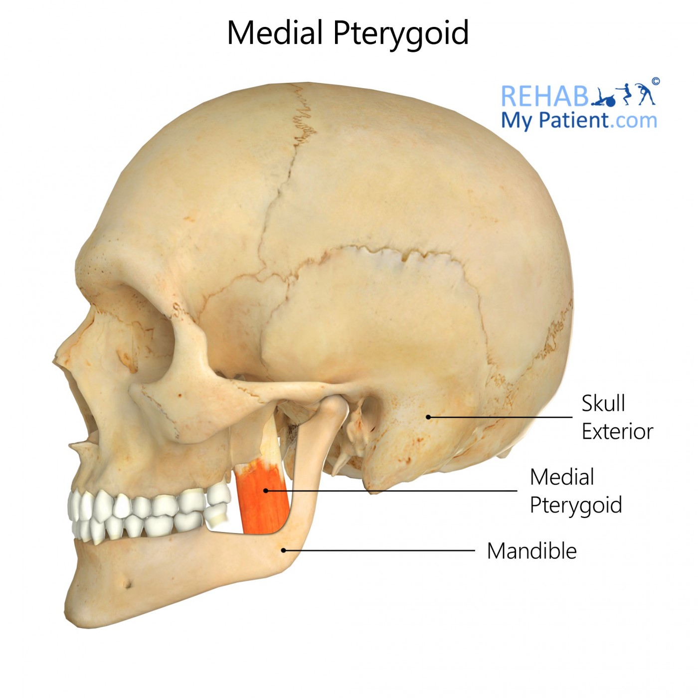
General information
The medial pterygoid works in combination with the masseter to aid in chewing.
Literal meaning
Wing-shaped – The muscle that attaches to the wing-shaped process of the sphenoid bone.
Interesting information
Medial pterygoid is a skeletal muscle located on either side of the jaw that assists in opening and closing of the mouth and with chewing.
Trismus, or “lockjaw”, is a condition where muscle spasms occur to the muscles that aid in mouth opening and closing. The condition is normally temporary but may be difficult to treat due to limited access to the oral cavity. Lockjaw can interfere with daily activities like eating and drinking, speaking, and oral hygiene. A number of contributing factors may play a role in the development of lockjaw including, Temporomandibular Joint Disorder, removal of wisdom teeth, mumps, or a needle prick to the medial pterygoid muscle. Treatment of the condition is dependent upon its severity. The use of NSAIDs, such as ibuprofen, and warm compresses to treat the pain and inflammation of the muscles, may help to ease the condition of lockjaw. For cases that do not improve with this treatment, the use of physical therapy or range of motion devices may rehabilitate the patient.
Origin
Deep head – Medial side of the lateral pterygoid plate behind the teeth.
Superficial head – Pyramidal process of the palatine bone and maxillary tuberosity.
Insertion
Medial angle of the mandible.
Function
Elevation of the mandible.
Closing of the jaw.
Side to side movement of the jaw.
Nerve supply
Mandibular nerve.
Blood supply
Pterygoid branches of the maxillary artery.
Relevant research
Heterotopic bone formation within a muscle, Myositis ossificans traumatica (MOT), often occurs to individuals who have experienced trauma to the area, repeated injury, burns, or surgical manipulation. A case study shows the development of rare bone formation within the medial pterygoid. Upon further investigation, the woman previously underwent a procedure to extract the lower third molar upon a dental visit. During the extraction, there was the necessity of several injections to achieve anaesthesia and to extract the tooth plus the mouth remained open for a prolonged period. Surgery tends to be the only treatment for cases of MOT. In this case, surgery was unable to fully remove the bone formation.
Thangavelu, A, Vaidhyanathan, A, Narendar, R. (2011). “Myositis ossificans traumatica of the medial pterygoid”. International Journal of Oral and Maxillofacial Surgery. 40:5, 545-549.
Medial pterygoid exercises
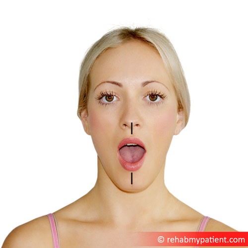
Open wider
Begin by slowly opening mouth as wide as possible. If clicking or popping of the jaw occurs, discontinue stretch immediately. Hold stretch for ten seconds then close jaw. Repeat this stretch ten times, three days a week.
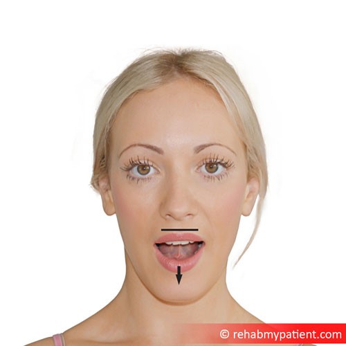
Tongue Stretches
Move the tongue to where it rests on the back of the upper front teeth. Next, move the tongue back until it is resting on the soft tissue of the roof of the mouth. Open the mouth, making sure to keep the tongue in the same position. Hold for ten seconds and return to the starting position. Complete three repetitions of these exercises two times a week.
Sign Up
Sign up for your free trial now!
Get started with Rehab My Patient today and revolutionize your exercise prescription process for effective rehabilitation.
Start Your 14-Day Free Trial