
General information
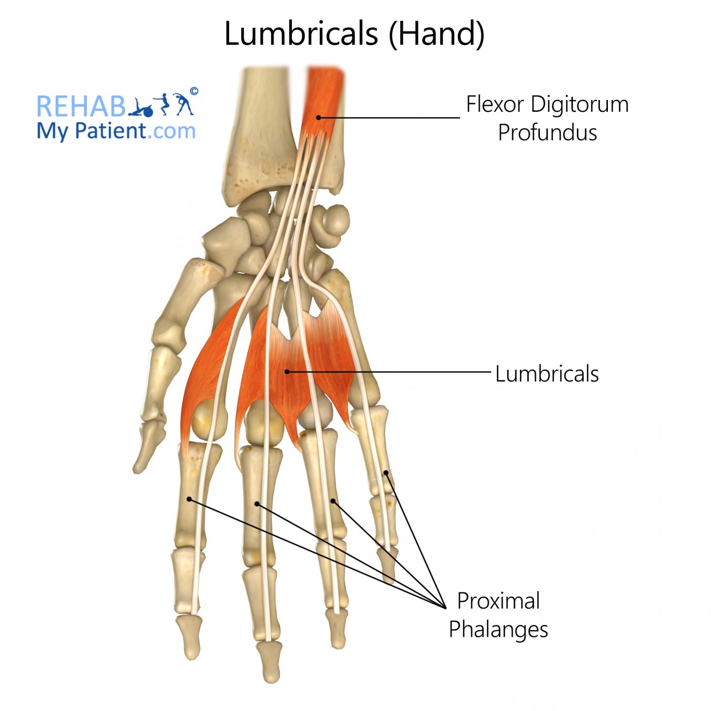
Lumbrical hand muscles are unique because they attach to tendons instead of bone. These tendons are the flexor digitorum profundus and the extensor expansions.
Literal meaning
Worm muscle.
Interesting information
The lumbricals are worm-like skeletal muscles located in the hand that aid in interchanging motion and fine digit-tuning motion.
The primary cause for tearing a lumbrical muscle of the hand is rock climbing. With unique hand positions, climbers are prone to supporting a full body’s worth of weight with two fingers. When the middle and ring finger remain in extension in a hold for an extended period of time, small tears or large rips can occur in the lumbrical. The use of alternate hand techniques and stretching the hand ahead of time are methods that can prevent the rip of the lumbrical muscle. Individuals should not treat lumbrical tears with anti-inflammatories. Treatments include rest of the muscle, physical therapy with ultrasound therapy, and stretching. A sonogram will determine the severity of the injury, which will determine the amount of time healing will take. Resumption of activities can take anywhere from ten to thirty days.
Origin
Flexor digitorum profundus.
Insertion
Extensor expansion.
Function
Flexion of metacarpophalangeal joints.
Extension of interphalangeal joints.
Nerve supply
3rd and 4th deep branch of ulnar nerve.
1st and 2nd median nerve.
Blood supply
Superficial palmar arch.
Common palmar digital artery.
Deep palmar arch.
Dorsal digital artery.
Relevant research
Assessments of the location of the lumbrical muscles (LM) in conjunction to the transverse carpal ligament explored the differences in carpal tunnel patients’ and cadavers’ LM locations. Differences in LM locations may determine that some individuals may be more prone to develop carpal tunnel syndrome. Before surgery, the conduction of examinations recorded the LM origins in relation to the transverse carpal ligament. The dissection of cadaver hands allowed for the comparisons between non-carpal tunnel syndrome hands versus hands with the carpal tunnel syndrome. Findings show that the location of the LM muscle from the carpal tunnel hands had a significantly different location than the cadaveric hands, with the LM origin located more proximally in the canal. Another observations showed that patients who performed repetitive motions with their hands exhibited larger lumbricals.
Siegel, D, Kuzma, G, Eakins, D. (1995). “Anatomic investigation of the role of the lumbrical muscles in carpal tunnel syndrome”. The Journal of Hand Surgery. 20(5), 860-863.
Lumbricals (hand) exercises
Finger stretch #1
Bend fingers down until all knuckles are bent. The tips of the fingers should touch the pads at the base of the fingers. Hold the stretch for a few seconds and repeat with the opposite hand. Complete the stretch five times daily.

Finger stretch #2
Bend fingers down so the tips of the fingers rest on the palm of the hand. Hold for a few seconds and repeat with opposite hand. Complete the stretch five times daily.
The “L” Stretch
Keeping the knuckles straight, bend fingers forward forming an L shape with the fingers and the hand. Hold for a few seconds and repeat with opposite hand. Complete the stretch five times.
Sign Up
Sign up for your free trial now!
Get started with Rehab My Patient today and revolutionize your exercise prescription process for effective rehabilitation.
Start Your 14-Day Free Trial