
General information
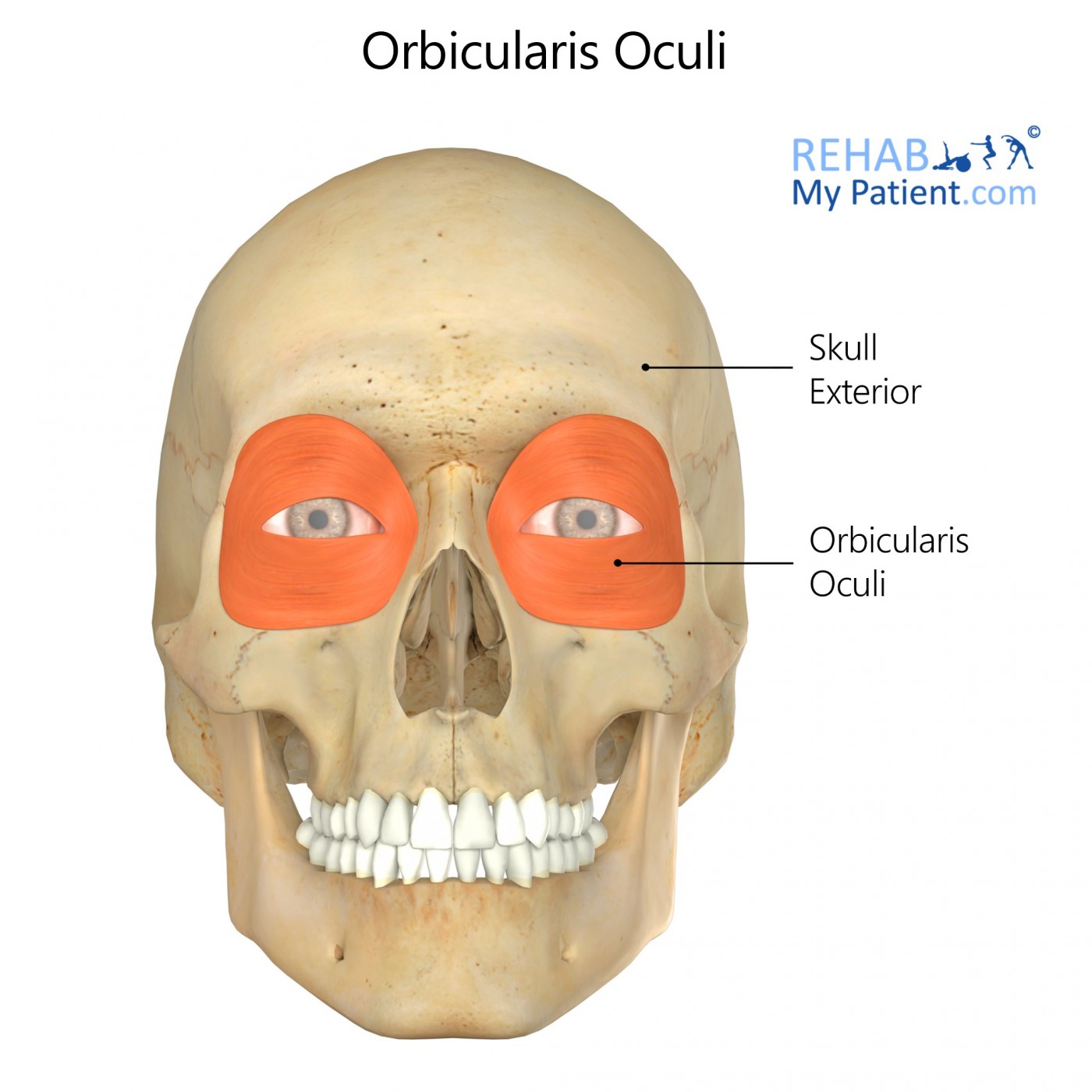
Orbicularis oculi is the muscle surrounding the eye.
Literal meaning
The little circle muscle of the eye.
Interesting information
Orbicularis oculi is a skeletal muscle of the face that surrounds the eye and is responsible for closing the eye.
Injury to the orbicularis oculi can result from overuse, which may result in headaches, eyestrain, or sinus headaches. The main cause for overuse of this muscle is poor eyesight without the proper corrective lenses. If eyesight is not corrected then the eyes strain in order to try to see better, which uses the orbicularis oculi. NSAIDs, like ibuprofen, will help with headaches and proper eyewear may prevent strain. If left untreated, the orbicularis oculi can cause crow’s feet to develop.
Origin
Frontal bone.
Medial palpebral ligament.
Lacrimal bone.
Insertion
Lateral palpebral raphe.
Function
Closes the eyelids.
Nerve supply
Temporal and zygomatic branches of the facial nerve.
Blood supply
Ophthalmic artery.
Zygomatico-orbital artery.
Angular artery.
Relevant research
Previously conducted eyelid surgeries used techniques based on the premise that the nerves to the orbicularis oculi muscle approach it from the lateral and that the segmental fascicles go parallel to the muscle fibres. Surgeries based off this premise have many issues including ectropion, the flipping of the eyelids so the inward part is exposed, and other incorrect eyelid positions. An anatomical study indicated that techniques using the approach through the lower eyelid might change the innervation of orbicularis oculi muscle. This innervation change would put the muscle at a much higher risk for complications.
Ramirez, O, Santamarina, R. (2000). “Spatial orientation of motor innervation to the lower orbicularis oculi muscle”. Aesthetic Surgery Journal. 20:2, 107-113.
Blepharoptosis occurs when the upper portion of the eyelid is less than 2.0 mm from the mid-pupil or there is more than 2.0 mm of asymmetry between the two eyelids. This condition has been a challenge for surgeons due to the complications that may arise from conventional surgeries. Due to previous research, the conclusion came to develop a Frontalis-Orbicularis Oculi Muscle (FOOM) flap-shortening technique. This technique allows for the harvesting of the FOOM flaps and the adjustment of the flaps depending on the severity of the condition. The procedure prevents the eye from closing or drooping involuntarily and enhances the ability to keep the eye wide open. This study showed that the procedure yielded good results with the biggest issue being under-correction.
Lai, C, Lai, C, Huang, S, Feng Sun, I, Chang, K, Lee, S, Lin, S. (2010). “A new trend for the treatment of blepharoptosis: Frontalis-Orbicularis Oculi Muscle flap shortening technique”. Journal of Plastic, Reconstructive & Aesthetic Surgery. 63:2, 233-239.
Orbicularis oculi exercises
The eyelid squeeze
Begin by placing the fingertips on the eyebrows. Close the eyelids and squeeze tightly. Hold the position for forty seconds. Practice this exercise five times a day.
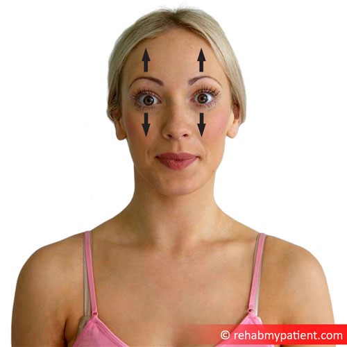
Surprise eyes
Begin sitting with good posture. Raise the eyebrows up as far as possible and open the eyes widely. Hold this position for ten seconds, release for five seconds, and then repeat ten times. Perform this exercise daily.
Sign Up
Sign up for your free trial now!
Get started with Rehab My Patient today and revolutionize your exercise prescription process for effective rehabilitation.
Start Your 14-Day Free Trial