
General information
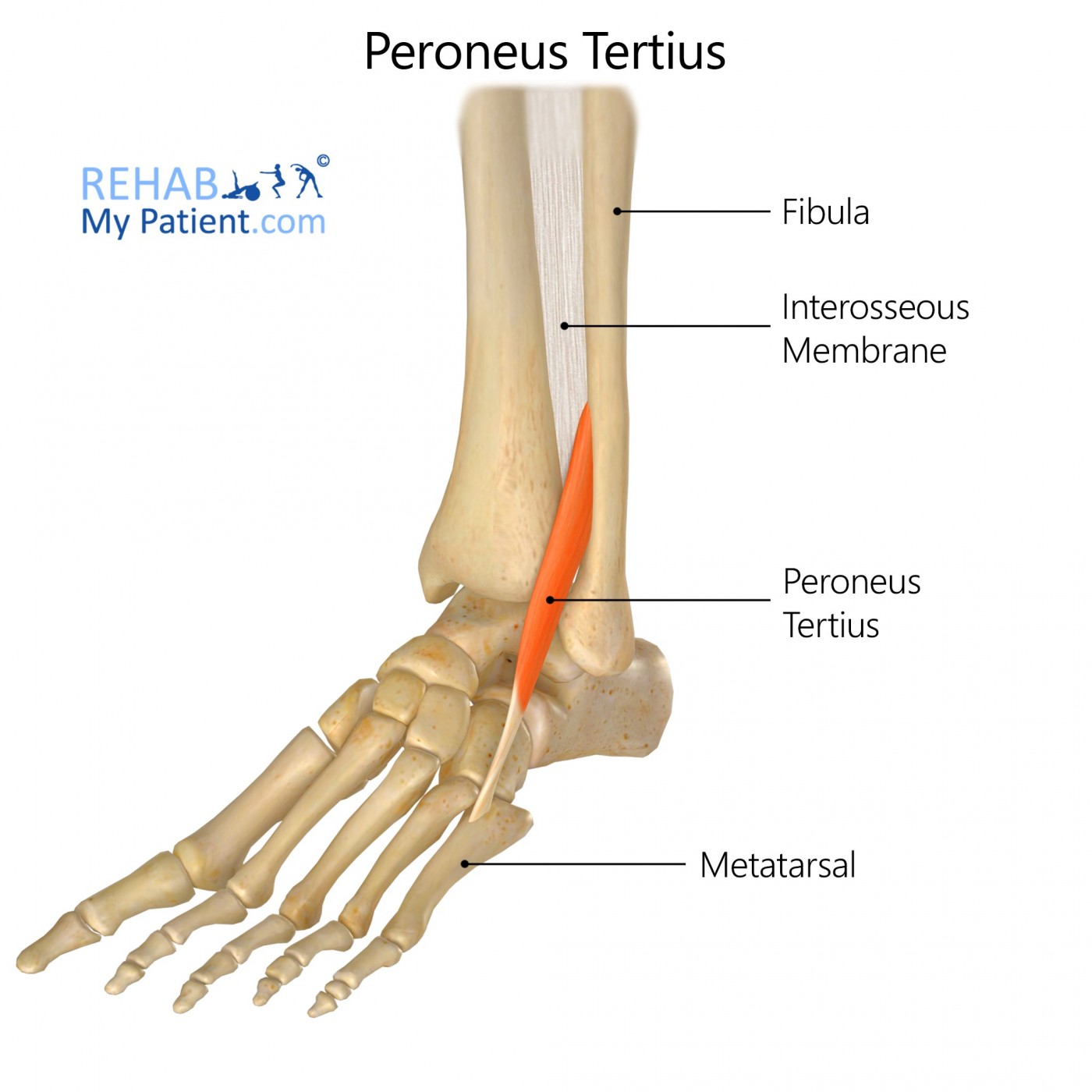
The peroneus tertius is a muscle found in the lower limb of the human body. It comes from the lower third portion of the fibula’s anterior surface, from the intermuscular septum and the lower portion of the interosseous membrane.
Literal meaning
The third muscle in the brooch, or fibula.
Interesting information
The peroneus tertius may be difficult to locate in some people. One out of every twelve people are born without this muscle. One in every seven people have an additional muscle here. At the bottom of this muscle, it attaches to the fourth and fifth metatarsals. If this muscle is injured, pain is located on the front of the anklebone and the outer part of the heel. To differentiate this pain from a sprain, check the severity of the pain. Sprains tend to be less severe than damaging tissues. Trouble raising the injured foot and weakness in the ankle are probably due to injury to the peroneus tertius muscle. NSAIDs, such as ibuprofen, and icing the area aid in reducing the pain and inflammation.
Origin
Medial surface for the distal third for the fibula and its adjacent interosseous membrane.
Insertion
Medial side on the surface of the dorsal at the base of the fifth metatarsal and into the shaft of the bone.
Function
Eversion of the foot.
Dorsal flexion in the joints of the ankle.
Nerve supply
Deep peroneal nerve S1 and L5.
Blood supply
The anterior tibial, anterior lateral malleolar, and fourth dorsal metacarpal arteries. Also from arcuate artery and peroneal artery perforating branches.
Relevant research
When the portals in the feet are not properly positioned, it can cause visualisation issues. This leads to problems being able to diagnose and treat the condition. Since there are potential serious complications with arthroscopy, the trans-Achilles and anterocentral portals are no longer used. Some of the other portals in the foot tend to provide easier access to the muscles, especially that of the posterior compartment of the ankle. Understanding the portals and structures in the ankle is imperative to repairing damage done.
Golano, Pau, Vega, Jordi, Perez-Carro, Luis, Gotzens, Victor (2006). Ankle Anatomy for the Anthroscopist. Part I: The Portals. Foot and Ankle Clinics. Vol. 11, Is. 2, 253-273.
Peroneus tertius exercises
Foot eversion
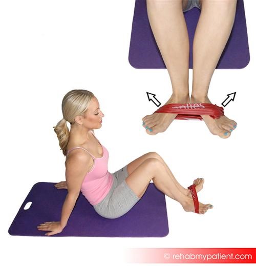
Tie a resistance band to something sturdy, such as the leg of a table. Sit on the floor with the legs straight out in front of the body, and position the foot into the loop at the ball of the foot and with the loop placed on the outside of the foot. Begin with the toes facing up to the ceiling, then turn the foot outward. Return the foot back to the beginning position, making sure to keep the rest of the leg as still as possible. Complete 10 to 20 repetitions on each side.
Calf raises
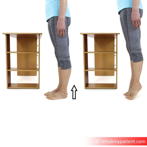
Begin standing with both feet flat on the floor at shoulder-width apart. Keep the rest of the body straight, but without locking the knees. Slowly, press on the toes, causing the heels to raise up off the ground. Hold this raised position for a couple of seconds, then slowly return the heels back to the ground. Perform ten repetitions for three sets. Additional resistance may be added by holding five-pound weights in each hand with the arms down beside the body while performing the exercise.
Sign Up
Sign up for your free trial now!
Get started with Rehab My Patient today and revolutionize your exercise prescription process for effective rehabilitation.
Start Your 14-Day Free Trial