
General information
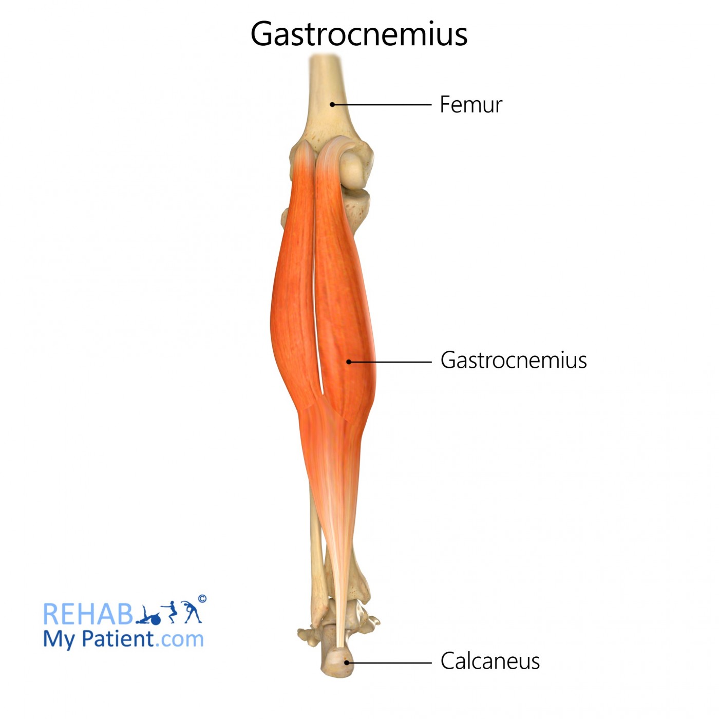
Gastrocnemius is a strong and prominent muscle of the lower leg. It is part of the calf muscle group.
Literal meaning
The belly muscle of the leg.
Interesting information
The name of gastrocnemius is derived from the Greek words for stomach and leg because the bulging mass of the muscle resembles a “belly of the leg”. Along with the soleus muscle, it comprises the calf muscle group. This powerful muscle runs from the back of the knee all the way down to the heel. It is absolutely essential for locomotion including walking, running, and jumping.
The origin of the muscle has both a lateral and medial head. The medial head is connected to the medial condyle of the femur bone while the lateral head connects at the lateral condyle of the femur. Gastrocnemius eventually joins with the soleus muscle to form the calcaneal tendon, also referred to as the Achilles tendon. Since gastrocnemius and soleus share a common insertion some anatomists consider them to be a single muscle.
Painful muscle spasms (involuntary contractions) are not uncommon in gastrocnemius and they can be influenced by a number of factors including dehydration and repetitive use. Gastrocnemius can also become inflamed from overuse. Anti-inflammatory medication such as ibuprofen along with a cool compress and elevation can be used to treat inflammation.
In rare cases, anatomical anomalies in gastrocnemius (the medial head in particular) may result in a condition called popliteal artery entrapment syndrome. This is a very serious condition that, if left untreated, can cause significant artery degradation, artery occlusion, or aneurysm.
Origin
Medial and lateral condyles of the femur.
Insertion
Posterior side of the calcaneus.
Function
Flexion (bending) of the knee as well as plantar flexion of the ankle and foot.
Nerve supply
Tibial nerve (S1, S2).
Blood supply
Sural arteries.
Relevant research
Recent trials in rodents have shown great potential for using ultrasound techniques to measure gastrocnemius muscle volume which can be correlated with muscle atrophy. This method is non-invasive and easy to perform and could be significant in assessing muscle atrophy after nerve damage.
Nijhuis THJ, de Boer SAS, Wahegaonkar AL, Bishop AT, Shin AY, et al. (2013). “A New Approach to Assess the Gastrocnemius Muscle Volume in Rodents Using Ultrasound; Comparison with the Gastrocnemius Muscle Index”. PLoS ONE 8(1): e54041. doi:10.1371/journal.pone.0054041.
Electromyography has shown that static stretching of gastrocnemius can actually decrease subsequent jumping performance, particularly vertical jump. This has important implications for physical therapists, athletes, and coaches.
Wallmann HW, Mercer JA, McWhorter JW. (2005). “Surface electromyographic assessment of the effect of static stretching of the gastrocnemius on vertical jump performance”. J Strength Cond Res. 19(3):684-8.
Gastrocnemius (leg) exercises
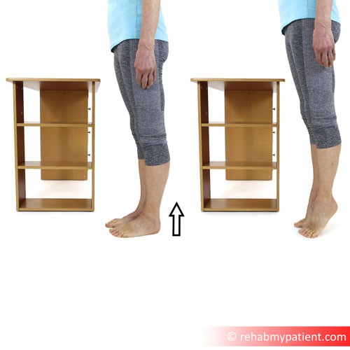
Standing calf raises are an excellent way to isolate and strengthen gastrocnemius. Stand tall with your feet, knees, hips, and shoulders nicely aligned. Your feet should be about shoulder width apart. Slowly shift your weight to the balls of your feet and use your calf muscles to lift your body weight. Make sure you keep your abdominal muscles tight and do not lean forward. Hold for 30 seconds then return to starting position. Repeat five times with ten seconds rest between sets.
To increase resistance you can hold dumbbells in your hands or try doing a single calf raise with your full body weight supported by only one leg at a time.
Sign Up
Sign up for your free trial now!
Get started with Rehab My Patient today and revolutionize your exercise prescription process for effective rehabilitation.
Start Your 14-Day Free Trial